
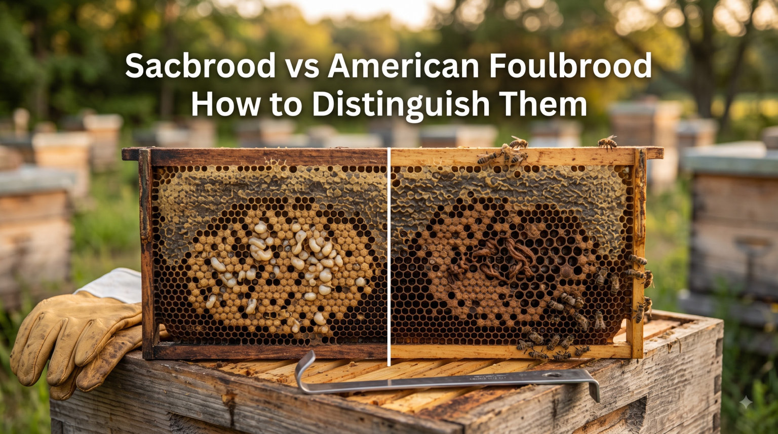
Opening a hive and spotting dead or discolored brood is one of the most unsettling moments a beekeeper can face. The question that follows is almost always the same: is this sacbrood, or is it something far more dangerous like American Foulbrood? Getting that answer right matters more than almost any other diagnosis in the apiary.
The Sacbrood vs American Foulbrood distinction is the difference between a manageable viral infection and a devastating bacterial disease that can wipe out an entire operation. American Foulbrood (AFB) is reportable in most countries, and confirmed cases often require colony destruction and equipment burning. Sacbrood, while still concerning, usually resolves on its own with good colony management.
In this guide, I walk you through every visual sign, diagnostic test, and field workflow you need to tell these two diseases apart with confidence. You will learn what each disease looks like inside the cell, how to perform the ropiness test correctly, and what to do the moment you suspect a problem. Everything here is based on established diagnostic protocols from agricultural biosecurity agencies and decades of beekeeping field experience.
Whether you are a new beekeeper inspecting your first few hives or an experienced apiarist managing dozens of colonies, this field guide gives you a clear, step-by-step process for brood disease identification. Let us start with the fastest way to tell these diseases apart.
Table of Contents
Sacbrood vs American Foulbrood: Quick Comparison
The quickest way to separate these two diseases comes down to three things: what the dead larva looks like, how it behaves when you probe it, and what the capping above it looks like. Here is the side-by-side field reference that most beekeeping guides fail to put in one place.
Affected stage: Sacbrood kills larvae in the late larval or prepupal stage, after the cell is capped. American Foulbrood kills larvae slightly earlier, typically at the late larval stage just before or after capping.
Dead larva appearance: Sacbrood larvae turn into a fluid-filled sac that eventually dries into a brittle, gondola-shaped scale that lifts out easily. AFB larvae decay into a wet, brown, sticky mass that dries into a hard scale tightly glued to the cell floor.
Ropiness test: Sacbrood remains do NOT produce a ropey thread. AFB remains produce a distinctive brown mucus-like thread that stretches at least 2 to 3 centimetres when pulled.
Smell: Sacbrood has little to no smell, sometimes faintly sour. AFB produces a strong, distinctive sulphurous or “foul” odour that experienced beekeepers recognize instantly.
Cappings: Sacbrood shows perforated, sunken, or greasy-looking cappings scattered across the brood nest. AFB shows the same signs but in a more concentrated, spreading pattern, often with a dark, sunken, and wet appearance.
Scale adhesion: Sacbrood scales are brittle and lift out of the cell easily with a matchstick or hive tool. AFB scales are firmly cemented to the lower cell wall and are extremely difficult to remove.
Cause: Sacbrood is caused by the Sacbrood virus (Iflavirus genus). American Foulbrood is caused by the spore-forming bacterium Paenibacillus larvae.
Severity: Sacbrood is generally mild and self-limiting. American Foulbrood is one of the most destructive honey bee diseases known and is legally notifiable in most regions.
Keep this comparison handy during hive inspections. Now let us examine each disease in detail so you understand exactly what you are looking at.
Understanding Sacbrood
What Causes Sacbrood?
Sacbrood is a viral disease caused by a pathogen in the Iflavirus genus, commonly called the Sacbrood virus. It is one of the most widespread honey bee viruses in the world, present in colonies on nearly every continent where beekeeping is practiced.
The virus infects larvae through contaminated food fed by nurse bees. Once inside the larva, the virus replicates rapidly and interferes with the moulting process, preventing the larva from pupating properly. The larva dies in the prepupal stage, still inside its unshed skin.
Unlike bacterial brood diseases, the sacbrood virus does not produce spores. This means it is less persistent in the environment than American Foulbrood, though it can still linger in wax, pollen stores, and on the bodies of adult bees. Colonies with strong hygienic behaviour often clear sacbrood infections on their own within a few weeks.
Sacbrood tends to appear during periods of stress, such as early spring buildup, nectar dearths, or when colonies are dealing with other pressures like Varroa mite infestations. Forum beekeepers frequently note that sacbrood is “common but not well recognized,” meaning many colonies carry the virus without showing obvious symptoms.
Visual Symptoms of Sacbrood
Recognizing sacbrood starts with knowing what healthy brood looks like. In a healthy colony, capped brood forms a solid, uniform pattern of evenly coloured cappings. Any break in that pattern deserves a closer look.
The first visual sign of sacbrood is a spotty or patchy brood pattern. You will see scattered uncapped cells or cells with perforated cappings mixed among healthy capped brood. The cappings may appear slightly sunken, darkened, or have a small hole where adult bees have attempted to clean out the dead larva.
When you open an affected cell, the dead larva is the key diagnostic feature. In sacbrood, the larva lies on its back in the cell, stretched lengthwise. It appears as a greyish to straw-coloured, fluid-filled sac. This sac is actually the unshed larval skin that has filled with fluid after death.
As the larva dries out over several days, it gradually changes colour from grey to dark brown or almost black. The dried remains take on a distinctive gondola or banana shape, curving along the bottom of the cell. This “banana shape” is one of the most reliable sacbrood field signs.
The final dried scale is brittle and flaky. Unlike AFB scales, sacbrood scales lift out of the cell easily with a matchstick or fine probe. Adult bees in a hygienic colony will often remove these scales themselves, which is why the overall brood pattern looks patchy rather than uniformly dead.
One important detail: sacbrood rarely produces a noticeable odour. If you open a cell and detect a strong foul smell, you are likely looking at a bacterial infection, not sacbrood.
Sacbrood Disease Cycle
The sacbrood disease cycle is relatively straightforward compared to bacterial brood diseases. Nurse bees inadvertently feed virus-contaminated brood food to young larvae during their first few days of life. The virus enters the larval gut and begins replicating.
The infected larva continues developing normally until it reaches the prepupal stage. At this point, usually one to two days after the cell is capped, the virus prevents the larva from completing its final moult. The larva cannot pupate and dies inside its unshed skin, which fills with fluid and forms the characteristic sac.
The dead larva remains in the capped cell unless adult bees detect and remove it. In colonies with strong hygienic behaviour, workers uncap the cell and drag out the sac, breaking the infection cycle. In colonies with weaker cleaning behaviour, the sac remains, dries into a scale, and continues producing virus particles.
Adult bees can also become infected with sacbrood virus without showing symptoms. These asymptomatic adult carriers spread the virus through glandular secretions, sharing food, and contact within the hive. This is why sacbrood can persist at low levels in apparently healthy colonies for long periods.
The entire cycle from infection to visible symptoms typically takes 5 to 8 days. This means that if you spot sacbrood today, the actual infection occurred over a week ago. Re-infection cycles can continue as long as conditions remain stressful for the colony.
Understanding American Foulbrood
What Causes American Foulbrood?
American Foulbrood is caused by Paenibacillus larvae, a gram-positive, spore-forming bacterium. This single-celled organism is one of the most resilient pathogens in the beekeeping world. The bacterial spores can survive for decades in wax, honey, and on contaminated equipment, even in extreme environmental conditions.
Only the spore form of the bacterium causes infection. When a young larva ingests spores through contaminated brood food, the spores germinate in the larval midgut. The vegetative bacteria then multiply rapidly, penetrating the gut wall and invading the larval body cavity.
The larvae are most susceptible to infection in their first 24 to 36 hours of life. After about 3 days of age, larvae become increasingly resistant. This is why AFB typically shows up in the youngest brood and why the disease advances gradually rather than killing an entire frame overnight.
The spores that cause AFB are extraordinarily persistent. They resist heat, cold, drying, and many disinfectants. An infected larva can produce up to a billion spores, and these spores remain viable in old comb and equipment for 50 years or more. This persistence is why AFB management often requires burning infected equipment rather than simply cleaning it.
American Foulbrood is classified as a statutory notifiable disease in the United States, the United Kingdom, Australia, Canada, and most other beekeeping nations. This means that suspected or confirmed cases must be reported to agricultural authorities, and control measures may be legally enforced.
Visual Symptoms of American Foulbrood
The visual signs of AFB develop as the infection progresses through the brood nest. The earliest and most reliable sign is a patchy, irregular brood pattern with scattered sunken and perforated cappings.
Unlike sacbrood, where affected cells are scattered loosely, AFB creates a more concentrated area of dead brood that gradually expands. The cappings over affected cells become sunken, dark, and sometimes moist or greasy in appearance. Many cappings develop small perforation holes where adult bees have attempted to investigate or clean the cell.
When you open an affected cell, the dead larva appears as a soft, moist, light-brown mass. Over time, this mass darkens to a rich coffee-brown colour and takes on a wet, glistening appearance. The consistency is sticky and paste-like, completely different from the fluid-filled sac of sacbrood.
The defining visual test is the ropiness test. When you insert a small stick or matchstick into the decaying mass and slowly withdraw it, the material forms a brown, mucus-like thread that stretches 2 to 3 centimetres or more before snapping. This ropy thread is the single most diagnostic sign of American Foulbrood.
As the larval remains continue to dry over several weeks, they form a hard, dark brown or black scale firmly attached to the lower cell wall. This scale is extremely difficult to remove. If you run a matchstick along the bottom of the cell, the AFB scale stays put, unlike a sacbrood scale which crumbles and lifts away.
The cappings over AFB-affected cells often show a distinctive “pepper pot” pattern when viewed at an angle. This pattern of scattered perforations across sunken cappings is a classic field sign that experienced inspectors look for during routine colony examinations.
AFB also produces a characteristic sulphurous, foul odour that gives the disease its name. Not every colony produces a noticeable smell, especially in early infections, but a strong foul odour combined with brown ropy larvae is essentially diagnostic.
American Foulbrood Disease Cycle
The AFB disease cycle begins when a young larva ingests Paenibacillus larvae spores with its brood food. The spores germinate in the midgut within hours, and the vegetative bacteria begin multiplying rapidly inside the larval digestive tract.
After 2 to 3 days of bacterial growth, the vegetative cells breach the gut wall and flood the larval body cavity. The larva dies shortly after, usually around the time the cell is capped or within the first day or two after capping. The bacteria continue digesting the larval tissues, reducing them to the characteristic brown, ropy mass.
Roughly 10 to 14 days after the larva dies, the bacterial population enters its sporulation phase. Millions of spores form inside the drying remains, creating the hard, dark scale on the cell floor. Each scale can contain over a billion spores, all capable of starting new infections.
Nurse bees attempting to clean out infected cells come into contact with these spores and spread them throughout the hive. They contaminate honey, pollen, wax, and their own mouthparts. When they feed other young larvae, the cycle repeats and the infection spreads to new cells and new frames.
The cycle is relentless without intervention. Because the spores are so persistent, even apparent recovery does not mean the disease is gone. Antibiotic treatment can suppress symptoms temporarily, but spores remain in the comb and equipment, ready to cause new outbreaks when the treatment stops.
This persistence is why agricultural authorities take AFB so seriously. A single infected colony can seed contamination across an entire apiary through robbing, drifting, and equipment sharing. The disease cycle, once established, is nearly impossible to break without destroying contaminated materials.
The Ropiness Test: A Step-by-Step Field Procedure
The ropiness test, also called the matchstick test, is the single most important field diagnostic for American Foulbrood. It takes less than a minute to perform and can distinguish AFB from sacbrood and other brood diseases on the spot.
Before performing the test, put on disposable gloves. If the result is positive for AFB, you will want to avoid spreading spores on your hands to other hives or equipment.
Step 1: Locate a suspect cell. Identify a cell with a sunken, perforated, or darkened capping. This is where dead or decaying brood is most likely to be found. Use your hive tool to gently remove the capping if it has not already been opened by the bees.
Step 2: Insert the probe. Take a clean matchstick, toothpick, or small twig and insert it gently into the cell. Push it down until it touches the larval remains at the bottom of the cell. You should feel it enter a soft, moist mass.
Step 3: Withdraw slowly. Pull the matchstick straight out of the cell very slowly, at a steady pace. Do not jerk it out. The slow withdrawal is what reveals the ropiness if AFB is present.
Step 4: Observe the thread. Watch what happens as the material leaves the cell. If AFB is present, a brown, mucus-like thread will stretch from the cell to the matchstick, extending 2 to 3 centimetres or more before snapping. This is a positive ropiness test.
Step 5: Interpret the result. If the material forms a long, elastic, brown thread, you have a presumptive positive for American Foulbrood. If the material breaks immediately, falls apart in flakes, or does not stretch at all, the result is negative, and you may be looking at sacbrood or another brood condition.
Step 6: Contain and disinfect. Regardless of the result, dispose of the used matchstick in a sealed bag. If the test was positive, change your gloves before touching any other hive. Clean your hive tool with alcohol or flame before using it again.
A negative ropiness test does not rule out AFB entirely, especially in very early or very late infections when the larval remains are too young or too dry to produce a thread. If you are still suspicious after a negative test, submit a sample to a diagnostic laboratory for confirmation.
Key Differences Between Sacbrood and American Foulbrood
Several critical differences separate these two diseases. Understanding each one gives you multiple diagnostic checkpoints, so you are never relying on a single sign.
Pathogen type: Sacbrood is caused by a virus (Iflavirus). American Foulbrood is caused by a spore-forming bacterium (Paenibacillus larvae). This fundamental difference drives every other distinction between them.
Death stage: Sacbrood larvae die in the prepupal stage, after the cell is capped but before pupation begins. AFB larvae die slightly earlier, usually at the late larval stage, around the time of capping or shortly after.
Larval appearance: Sacbrood produces a fluid-filled sac that dries into a brittle, gondola-shaped scale. AFB produces a wet, brown, paste-like mass that dries into a hard, dark scale firmly cemented to the cell wall.
Scale removal: Sacbrood scales lift out easily. AFB scales are nearly impossible to remove from the cell and stay firmly attached to the lower cell wall even when probed.
Ropiness: Only AFB produces the characteristic brown, ropy thread. Sacbrood never produces ropiness. This is the most reliable single test for distinguishing them in the field.
Odour: AFB produces a sulphurous, foul smell in moderate to advanced infections. Sacbrood produces little to no odour.
Pattern severity: Sacbrood typically appears as scattered individual affected cells within an otherwise healthy-looking brood nest. AFB tends to cluster in expanding patches that gradually consume the brood area.
Colony impact: Sacbrood rarely causes significant colony weakening and often resolves without intervention. AFB progressively weakens and eventually kills the colony, and the spores contaminate equipment permanently.
Persistence: Sacbrood virus is relatively fragile and does not persist for years in equipment. AFB spores can survive for decades in wax, honey, and on woodenware, making reinfection a constant threat.
Reporting requirements: Sacbrood is not reportable in most regions. AFB is a notifiable disease throughout most of the world, and confirmed cases trigger legally mandated control measures.
Step-by-Step Field Diagnosis Workflow
When you spot something unusual in the brood nest, having a systematic workflow prevents mistakes. Follow these steps in order during every inspection where you suspect a brood disease.
Step 1: Assess the overall brood pattern. Before opening any cells, look at the capped brood as a whole. Is the pattern solid and uniform, or patchy with scattered gaps? A healthy colony has a compact, even brood pattern. Scattered gaps are your first warning sign.
Step 2: Examine the cappings. Look closely for sunken, perforated, darkened, or greasy-looking cappings. Note whether the affected cappings are scattered randomly or clustered in patches. Scattered cappings suggest sacbrood or early EFB. Clustered, expanding patches of affected cappings raise concern for AFB.
Step 3: Open a suspect cell. Choose a cell with a perforated or sunken capping and carefully open it with your hive tool. Observe the colour, texture, and moisture of the remains inside.
Step 4: Note the colour and consistency. A greyish, fluid-filled sac points to sacbrood. A soft, brown, moist, paste-like mass points toward AFB. A white, chalky, mummy-like mass indicates chalkbrood. Yellowish, twisted, coiled larvae may indicate European Foulbrood.
Step 5: Perform the ropiness test. Insert a matchstick into the remains and withdraw slowly. A brown thread stretching 2 to 3 centimetres confirms presumptive AFB. No ropiness points away from AFB.
Step 6: Check scale adhesion. Try to lift the dried remains with a probe or matchstick. If the scale lifts out easily and crumbles, it is likely sacbrood. If it is glued firmly to the cell floor and resists removal, suspect AFB.
Step 7: Smell the cell. Bring the open cell close and take a careful sniff. A sulphurous, foul odour supports an AFB diagnosis. Little or no smell favours sacbrood.
Step 8: Check multiple cells. Never diagnose from a single cell. Open 5 to 10 suspect cells across different frames to confirm the pattern is consistent.
Step 9: Take action based on findings. If signs point to sacbrood, monitor the colony and improve conditions. If signs point to AFB, stop inspecting immediately, close the hive, disinfect your tools, change gloves, and contact your local agricultural authority or bee inspector before doing anything else.
Step 10: Send a sample for confirmation. Even experienced beekeepers can misdiagnose brood diseases. If you have any doubt, collect a sample of the affected comb and submit it to an approved diagnostic laboratory for microscopic or molecular confirmation.
How Do These Compare to European Foulbrood and Chalkbrood?
Sacbrood and AFB are not the only brood diseases you might encounter. European Foulbrood (EFB) and chalkbrood are two additional conditions that can mimic these diseases and cause confusion during inspection.
European Foulbrood is caused by the bacterium Melissococcus plutonius. Unlike AFB, EFB affects larvae before they are capped. Infected larvae appear yellowish, twisted, and melted in their cells, often in a coiled or irregular position. They lose their normal pearly white colour and take on a creamy or yellow-brown tone.
EFB does not produce ropiness, which helps distinguish it from AFB. It also does not produce the hard, cemented scale characteristic of AFB. The dead larvae dry into a loose, rubbery scale that is easier to remove. EFB has a sour rather than sulphurous smell and is a notifiable disease in many regions, though its management differs from AFB.
Chalkbrood is caused by the fungus Ascosphaera apis. Infected larvae dry into hard, chalk-white, mummy-like remains that are easy to spot. These mummies may be white, or they may turn grey or black as fungal spores develop. Workers often drag chalkbrood mummies out of the hive, so you may find them on the landing board or in front of the entrance.
Chalkbrood mummies are dry and firm, completely different from the wet, ropy remains of AFB or the fluid-filled sacs of sacbrood. If you can pick up the dead brood and it feels like a piece of chalk, you are dealing with chalkbrood, not a bacterial or viral disease.
The key to distinguishing all four diseases lies in the stage of larval death, the appearance and consistency of the remains, and the results of the ropiness test. When in doubt, always confirm with a laboratory diagnosis.
How These Brood Diseases Spread
Understanding how brood diseases move between colonies helps you protect your apiary. Both sacbrood and AFB spread through similar pathways, but the risks differ dramatically because of the spore issue.
The primary transmission routes include contaminated equipment shared between hives, drifting bees entering the wrong colony, robbing of weak or dead colonies by strong ones, and swarming. Beekeepers themselves are a major vector through transferring infected combs, using contaminated tools, or feeding contaminated honey.
AFB is far more dangerous in its spread because the spores persist for decades. A single contaminated frame placed in a healthy hive can start an infection that destroys the colony months later. Sacbrood virus, by contrast, breaks down more quickly in the environment and poses a lower long-term contamination risk.
Purchasing used equipment, nucs, or package bees from unverified sources is one of the most common ways these diseases enter an apiary. Always inspect purchased colonies and quarantine new arrivals before integrating them with your existing stocks.
Management and Treatment Approaches
The management path for sacbrood versus American Foulbrood could not be more different. Getting this right protects not just your own colonies but those of every beekeeper in your area.
For sacbrood, the primary approach is supportive. Strengthen the colony by making sure it has adequate nutrition, a productive queen, and low Varroa mite levels. Requeening with a queen from a line known for hygienic behaviour can help, as these colonies are better at detecting and removing infected larvae. Most sacbrood outbreaks resolve within a few weeks without any specific treatment.
For American Foulbrood, management is strict and regulated. Suspected cases must be reported to your local bee inspector or agricultural authority immediately. Do not move combs, bees, or equipment from the affected hive. In many jurisdictions, confirmed AFB requires destruction of the colony and burning of all comb and woodenware. Some regions allow antibiotic treatment under veterinary supervision, but this suppresses symptoms without eliminating spores, so it is not a long-term solution.
The most effective AFB prevention strategy is never sharing equipment between hives, maintaining an apiary hygiene protocol, regularly inspecting brood, and participating in any voluntary or mandatory AFB monitoring programs in your region.
Common Beekeeping Mistakes When Diagnosing Brood Diseases
Beekeepers on forums like Beesource and Reddit consistently report the same diagnostic pitfalls. Learning from these mistakes can save your colonies and your confidence.
Mistake 1: Diagnosing from a single cell. One dead larva does not tell you what disease you are dealing with. Always check multiple cells across several frames before drawing any conclusions.
Mistake 2: Skipping the ropiness test. Many beekeepers identify diseases purely by appearance and miss the single most diagnostic test for AFB. Always carry matchsticks or toothpicks in your kit.
Mistake 3: Assuming mild symptoms mean mild disease. Early-stage AFB can look deceptively similar to sacbrood. A single perforated capping with slightly off-colour brood could be the first sign of a devastating infection.
Mistake 4: Not reporting suspected AFB. Some beekeepers delay reporting because they fear losing their colonies. Delaying only makes the situation worse, as the infection spreads to other hives and apiaries.
Mistake 5: Using contaminated tools across multiple hives. Hive tools, gloves, and even smoker bellows can transfer spores between colonies. Clean and disinfect your equipment between apiary visits.
Mistake 6: Confusing sacbrood with AFB and treating unnecessarily. If you misdiagnose sacbrood as AFB, you may destroy colonies and equipment that could have recovered with simple management improvements. Always confirm with a laboratory test before taking irreversible action.
FAQs
How do you tell if you have American foulbrood?
Look for a patchy brood pattern with sunken, perforated, and darkened cappings. Open suspect cells and check for brown, moist, paste-like larval remains. Perform the ropiness test by inserting a matchstick and withdrawing slowly. A brown thread stretching 2 to 3 centimetres confirms presumptive AFB. A sulphurous odour and hard, dark scales glued to the cell wall are additional signs.
How to identify EFB?
European Foulbrood affects uncapped larvae, causing them to appear yellowish, twisted, and melted in their cells. Infected larvae lose their pearly white colour and take on a creamy or brown tone. EFB does not produce ropiness and has a sour rather than foul smell. The dead larvae dry into a loose, rubbery scale rather than a hard, cemented one.
What does sacbrood look like?
Sacbrood appears as scattered cells with perforated or sunken cappings. Inside, the dead larva forms a greyish, fluid-filled sac lying on its back in the cell. As it dries, it turns dark brown or black and takes a distinctive gondola or banana shape. The dried scale is brittle and lifts out of the cell easily. Sacbrood produces little to no odour.
What is the difference between American and European foulbrood?
American Foulbrood is caused by Paenibacillus larvae and affects larvae around the capping stage, producing brown ropy remains and hard scales cemented to the cell wall. European Foulbrood is caused by Melissococcus plutonius and affects younger, uncapped larvae, producing yellowish, twisted, melted larvae without ropiness. AFB spores persist for decades, while EFB is less persistent in the environment.
Is American foulbrood harmful to humans?
No, American Foulbrood does not infect humans. Paenibacillus larvae only affects honey bee larvae. Honey from AFB-infected colonies is safe for human consumption, though it should not be fed to other bees as it contains infectious spores. The danger of AFB is entirely to bee colonies and beekeeping equipment.
Conclusion
Distinguishing Sacbrood vs American Foulbrood comes down to understanding the cause, appearance, and behaviour of the affected brood. Sacbrood is a viral disease that produces fluid-filled sacs and brittle, banana-shaped scales with no ropiness and little odour. American Foulbrood is a bacterial disease that produces brown, ropy masses, hard cemented scales, a sulphurous smell, and devastating colony losses.
Use the ropiness test as your primary field diagnostic, check multiple cells before concluding, and always confirm uncertain cases with a laboratory. If you suspect AFB, report it immediately and protect your neighbouring colonies by disinfecting equipment and avoiding any movement of bees or comb from the affected hive.
Keep this field guide with you during inspections, and revisit the step-by-step diagnosis workflow every time you spot something unusual in the brood nest. Early, accurate identification is the most powerful tool you have for protecting your bees in 2026 and beyond.